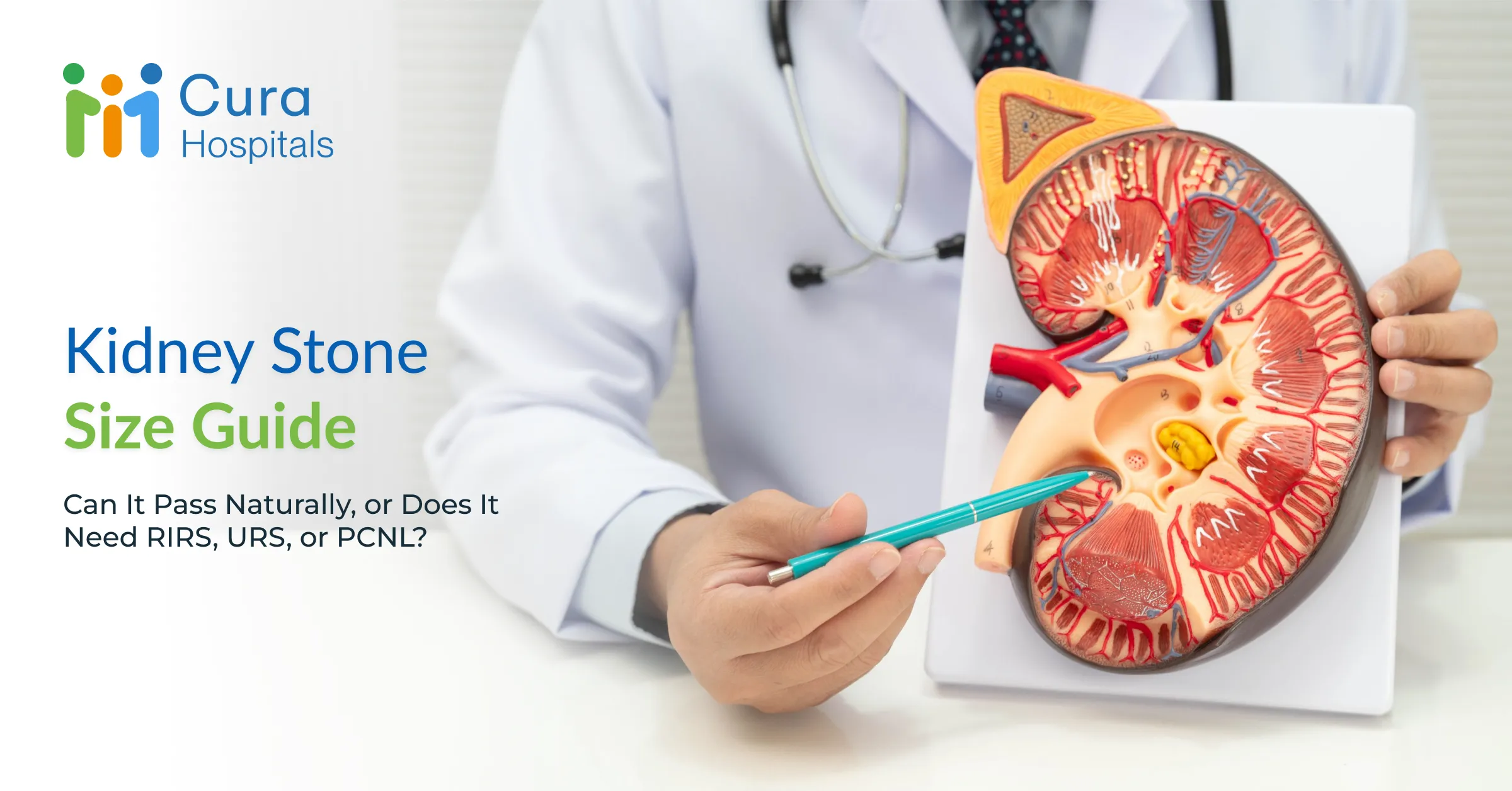
Kidney Stone Size Guide: Can It Pass Naturally, or Does It Need RIRS, URS, or PCNL?
 Cura Hospitals

When a sharp, sudden pain strikes your lower back or side, a diagnostic scan often confirms what you may have already feared: a kidney stone. Shortly after the report comes back, your doctor will mention a specific measurement, 4 mm, 7 mm, 12 mm, or larger.
Naturally, the first question on your mind is simple. Will it pass on its own, or will surgery be needed?
Stone size is a useful starting point, but choosing the right kidney stone treatment isn’t based on a single number alone. Where the stone is lodged, whether it’s blocking urine flow, and how dense it is all play an equally important role.
Here’s a clear guide to understanding kidney stone sizes, how urologists decide on the safest procedure for kidney stone removal, and what minimally invasive options are available at Cura.
Urgent Symptoms Needing Emergency Care
Most small kidney stones can be managed comfortably at home with fluids and pain medication. Certain warning signs, though, point to a medical emergency where a blocked stone is causing dangerous pressure or a spreading infection.
Seek immediate emergency care if your kidney stone comes with:
- Fever and chills. A high temperature alongside flank pain suggests an infected, obstructed kidney, a condition that needs urgent decompression.
- Uncontrollable pain or persistent vomiting. Severe, agonising pain that doesn’t ease with oral painkillers, or nausea intense enough that you can’t keep fluids down.
- Anuria, or an inability to pass urine. Passing no urine at all, or a sharp drop in output, is especially concerning if you have only one functioning kidney.
- Visible blood or pus in urine. Urine that looks thick, cloudy, or heavily tinged with dark blood.
Why Stone Size Is Only One Decision Factor
It’s tempting to assume a 5 mm stone automatically passes and a 9 mm stone automatically needs surgery. In reality, size is only one piece of the puzzle. Urologists weigh a whole set of physical and structural factors before recommending treatment.
A 6 mm stone floating freely inside a spacious kidney pocket causes no immediate blockage, while a 6 mm stone stuck in the narrow ureter, the tube connecting kidney to bladder, can cause severe obstruction. Urinary anatomy matters too: narrow passageways, congenital variations, or old scarring can make it hard for even tiny stones to travel out on their own.
Composition and toughness play a role as well, since calcium oxalate monohydrate or uric acid stones behave quite differently under treatment compared to softer struvite stones. And whether there’s infection or blockage present changes the urgency altogether. A small stone causing significant kidney swelling needs faster action than a larger, silent stone that isn’t obstructing urine flow at all.
Small, Medium, and Larger Stone Scenarios
Individual anatomy varies, but stone size still gives a useful general framework for what to expect.
| Stone Size | Likelihood of Natural Passage | Typical Recommended Approach |
| Less than 4 mm (very small) | High, around 80% or more | Conservative management: high fluid intake, pain relief, and observation |
| 4 mm to 6 mm (small) | Moderate, around 50 to 60% | Medical Expulsive Therapy (MET) with close monitoring; URS or RIRS if the stone gets stuck |
| 7 mm to 10 mm (medium) | Low, around 20% | Minimally invasive intervention preferred (URS or RIRS) |
| 10 mm to 20 mm (large) | Extremely low | RIRS or ESWL (shockwave lithotripsy), depending on hardness and location |
| Over 20 mm or complex (very large) | Negligible | Keyhole surgical removal (PCNL) |
This is also where a common question comes up: can you pass a 7mm kidney stone naturally? It’s possible, but the odds drop considerably at this size, which is why minimally invasive procedures are usually recommended rather than waiting it out.
4. Location and Obstruction: Why Where It Sits Matters
A stone’s position inside the urinary tract shapes both how bad the symptoms feel and which treatment path makes sense.
[Upper/Middle Kidney]──> Often asymptomatic until large ──> RIRS or PCNL
│
[Lower Pole Kidney] ──> Harder to flush out due to gravity ──> RIRS or PCNL
│
[Ureter (Narrow Tube)] ──> High risk of blockage/colic ──> URS or RIRS
│
[Bladder / Urethra] ──> Pain during urination / blockage ──> Cystoscopy / URS
Stones sitting quietly inside the kidney, in the renal pelvis or calyces, may cause only a dull ache or go unnoticed until they grow larger. Lower-pole stones tend to be harder to pass naturally, since they sit at the bottom of the kidney, working against gravity.
In the ureter, a narrow tube roughly 3 to 4 mm wide actively squeezes to move urine, a stone entering that tight passage triggers muscle spasms, producing the intense, wave-like pain known as renal colic. Stones also tend to get trapped at natural narrowings, such as the ureteropelvic junction or the vesicoureteric junction near the bladder.
Observation and Medical Expulsive Therapy (MET)
If a stone is under 6 mm, sits in a reasonably favourable position, and isn’t causing significant swelling or infection, your doctor may suggest a watchful waiting approach known as a trial of passage.
Conservative management usually involves drinking 2.5 to 3 litres of water daily to keep urine flow strong and help push the stone along. Pain is managed with NSAIDs or prescribed analgesics to control the muscle spasms. Your urologist may also prescribe medical expulsive therapy, commonly an alpha-blocker such as Tamsulosin, which relaxes the smooth muscle in the lower ureter, widening the channel and making the stone’s passage noticeably easier and less painful.
A trial of passage typically runs 2 to 4 weeks. If the stone hasn’t moved by then, or if the pain becomes unmanageable, a procedural intervention gets scheduled.
Surgical Options: URS vs. RIRS vs. PCNL
When a stone is too large, too hard, or firmly stuck, modern urology offers safe, effective procedures for kidney stone removal. Nearly all of today’s techniques are minimally invasive or endoscopic, meaning no large open incisions are involved.
- Ureteroscopy (URS)
Works best for medium to large stones sitting in the mid-to-lower ureter. A thin, rigid or semi-rigid scope passes through the natural urinary opening, through the urethra and bladder, directly into the ureter. A holmium or thulium laser breaks the stone into fine, dust-like fragments, which are then removed with a miniature basket. No incision is needed at all.
- Retrograde Intrarenal Surgery (RIRS)
Suits stones up to 20 mm sitting high inside the kidney or in the upper ureter. It uses a highly flexible, ultra-thin digital ureteroscope that can bend backward into the kidney’s inner chambers. A laser breaks the stone into dust, which then washes out naturally in the urine. Like URS, there’s no incision, and clearance rates tend to be excellent with minimal downtime.
- Percutaneous Nephrolithotomy (PCNL)
Reserved for large, complex, or staghorn stones over 20 mm sitting inside the kidney. Under general anaesthesia, a tiny keyhole track, about 1 cm, is created through the skin of the back directly into the kidney. A nephroscope goes in, and ultrasound or laser energy breaks up the stone so larger fragments can be removed right away. It leaves a single small puncture on the back and clears a heavy stone burden effectively in one sitting.
Diagnostic Tests and Recovery Considerations
Before deciding on the right intervention, your urologist will usually order a few specific scans to map the stone precisely.
A non-contrast CT scan (KUB) remains the gold standard for kidney stone evaluation, showing exact dimensions, location, density, and how much the kidney has swollen. An ultrasound (USG KUB) is radiation-free and useful for pregnant patients or for monitoring kidney swelling. A urine culture and routine test checks for any underlying infection that needs treating before a procedure, and kidney function tests, serum creatinine and BUN, confirm the kidneys are filtering normally.
[URS / RIRS] → Day care or 24-hr stay → Stent removed in 1-2 weeks → Full recovery: 2 to 5 days
[PCNL] → 1 to 2 days hospital stay → Minor back soreness → Full recovery: 7 to 10 days
A temporary thin plastic tube, called a JJ stent, is often placed inside the ureter after URS, RIRS, or PCNL to prevent swelling and keep urine draining smoothly. It comes out easily in a quick outpatient visit a week or two later. Most patients return to desk work within 2 to 4 days after RIRS or URS, while recovery from PCNL usually takes about a week.
For anyone weighing kidney stone size for surgery against simply waiting it out, this recovery picture is often the deciding factor, since the minimally invasive options come with a fairly quick return to normal life.
Urology Consultation at Cura
Living with ongoing kidney stone pain, or waiting endlessly to see if a stone passes, doesn’t have to be the only option. At Cura Multispeciality Hospitals, our urology team uses advanced diagnostic imaging along with minimally invasive laser technology, including laser RIRS and Mini-PCNL, to resolve kidney stones quickly, safely, and with as little disruption as possible.
Whether you need guidance managing a small stone at home or precise laser treatment for a complex one, our expert team is here to support your recovery at every step.
Schedule Your Urology Consultation at Cura Hospitals
Call Us: +91 87480 30303 | 24/7 Emergency: +91 84312 41403
Locations: Kammanahalli & Kanakapura Road, Bengaluru
Media Center Health Concerns?
Our specialists at Cura Hospitals are here to help with screening, diagnosis, risk assessment, and personalized treatment plans tailored to your health needs.
Schedule A ConsultationFrequently Asked Questions
Neither is strictly better; they target different areas. URS is best for stones stuck lower in the ureter, while RIRS uses a flexible scope to reach stones deep inside the kidney.
PCNL is typically required for large or complex stones greater than 20 mm (2 cm) that are too big to clear using laser procedures.
RIRS is generally more expensive due to the use of delicate flexible scopes, advanced laser equipment, and specialized single-use tools.
No. The ureter is only 3–4 mm wide, making natural passage virtually impossible and posing a high risk of blockage and kidney strain.
Generally, no. Most stones are made of calcium and cannot be dissolved. Only rare, pure uric acid stones might respond to medication, but a 15 mm stone almost always needs removal.
Related Blogs

Leg Pain, Swelling or a Non-Healing Wound: Vascular Surgeon, Orthopaedic Doctor or Diabetologist?

Your Health Check Report Is Abnormal. Which Doctor Should You Consult Next?